


Filters

Stock royalty-free photos and images of Naskórka
Discover unlimited high resolution images of Naskórka and stock visuals for commercial use.

The human skin is the outer covering of the body. In humans, it is the largest organ of the integumentary system. The skin has multiple layers of ectodermal tissue and guards the underlying muscles, bones, ligaments and internal organs.
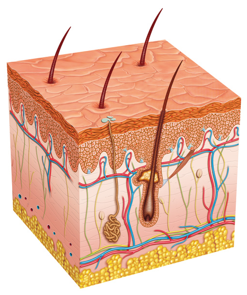
Image you can see the different layers and elements of the skin

Schematic illustration of the skin. It provides protection and isolation of the environmental organism. There are three main layers that theThey make up: epidermis, dermis, subcutaneous tissue and skin accessories such as hair, sebaceous glands and

Isometric hair growth cycle, anagen, telogen, catagen phases. Human skin layers with hair follicle growth flat vector illustration set. Hair growth stages

Cut of tanned skin, with surface of the skin, hairs and the sebaceous gland.

Skin with acne. 3d illustration of the structure of the skin layers microstructure.Medical and educational pictures isolated. High quality photo

Cut of skin and a hair with the sebaceous gland. .

Hair in the skin with the sebaceous gland and the horripilator muscle.

3d renderings of skin anatomy

Skin with wrinkles and age spots. 3d illustration of the structure of the skin layers microstructure.Medical and educational pictures isolated. High quality photo
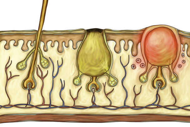
Histology of acne lesions. Illustration showing normal hair with sebaceous glands (left), formation of open comedone (center) and inflammatory papule (right)

Human skin. Layered epidermis with hair follicle, sweat and sebaceous glands. Healthy skin anatomy medical vector illustration. Dermis and epidermis skin, hypodermis, flat design

Realistic human dry skin cross-section of layers 3d isolated medical illuatration. Cracked skin. High quality 3d illustration

Cut of skin with hair (integument), epidermis, sebaceous gland. .

Human skin cutaway diagram, with several details. 2D digital illustration with clipping path, on white background.

Diagram of human skin structure

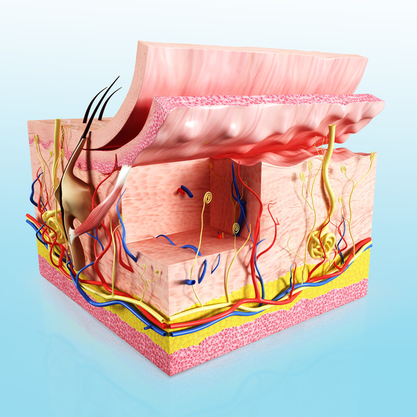
3d rendered illustration of front side of tissue repair of human skin
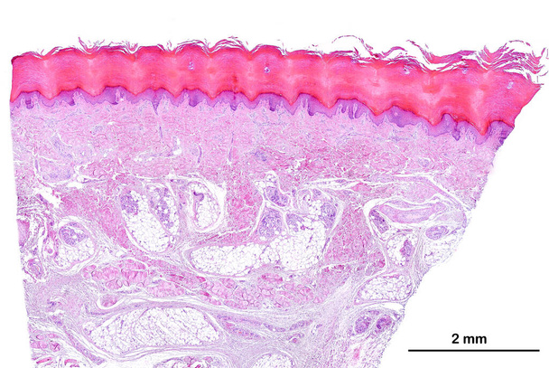
Low power light microscope micrograph of human glabrous skin. From top, the epidermis, a keratinized stratified squamous epithelium showing a prominent horny layer, the dermis and hypodermis. The hypodermis is identified by the presence of adipose ti

Inflammation is a process by which white blood cells protect of the body from infection with bacteria and viruses

Skin anatomy. Human body skin illustration with parts vein artery hair sweat gland epidermis dermis and hypodermis. Human Cross-section of the skin layers structure

Processes in derma

Dark skin type with lots of melanin grains.
3d rendered illustration of Human skin cut way diagram

The anatomical structure of the skin. Skin aging

Image of a part of the skin affected by the fungus

Scalp and hair follicles of human under the microscope in Lab.

Water circulation in the plant root, stem and leaf

Structure of the human skin. Layers and cells, isolated white.
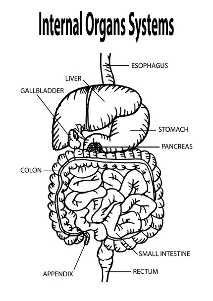
Human internal organs. Hand drawing illustration.

The anatomical structure of the skin. Elastin, hyaluronic acid, collagen. Skin aging, wrinkles before and after

Plant stem with sieve cells under the microscope.

The process of engraftment of the pigment after the salon procedure of permanent make-up. Illustration

3d illustration of human skin with hairs, microscopic view of skin, microscopic view of hairs

Affected skin of a child's corrosion dry crust flakes for medicine

Section of skin with the layers of the epidermis.

Educational model human skin tissue isolated on white background

Cross section through cells of a root from a maize plant (Zea mays) under the microscope.

Meso thread Lift. Young female with clean fresh skin. Beautiful woman. face and neck. Lifting by threads concept. Collage

Plant cell surface of leaf under light microscope
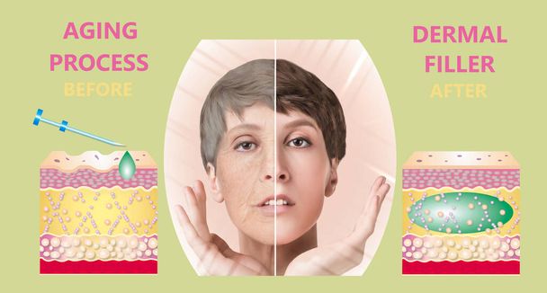
Cosmetic or dermal fillers. Young female with clean fresh skin. Beautiful woman. face and neck. Lifting by filler concept. Collage. Before and after

Cosmetic or dermal fillers. Young female with clean fresh skin. Beautiful woman. face and neck. Lifting by filler concept. Collage. Before and after

Hyaluronic acid. Skin-care products. The rejuvenation with help of treatment. Cosmetic or dermal fillers. Young female with clean fresh skin. Beautiful woman. face and neck. Lifting by filler concept

Section of the epidermis of white skin with few grains of melanin.

Acne vulgaris in an overweight person and closeup view of bacteria Cutibacterium acnes, formely formerly Propionibacterium acnes, associated with acne development, 3D illustration

Dangerous sunburn. The shoulders of a young girl.

Pink plant cells under microscope.

Peeling of skin after burned by sunlight. not using sunscreen. sunburn of the skin

Pain regulation: pain information can be bypassed by sensations of touch.

Cells of a plant leaf with damaged epidermis and chloroplasts under a microscope. The damage is caused by a disease.
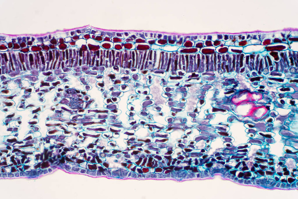
Cross section leaf of Plant under the microscope view for education.

Simple columnar epithelium is a columnar epithelium that is uni-layered. In humans, a simple columnar epithelium lines most organs of the digestive tract including the stomach, small intestine, and large intestine.

2d cartoon illustration of skin anatomy

ATP synthase 3d illustration

Cross section through cells of a root from a maize plant (Zea mays) under the microscope.

Photo of healthy humans skin, macro shooting

Microscope photo of the cells from the bark from a cork tree.

Shingles is a viral infection that causes a painful rash. shingles can occur anywhere on your body, it most often appears as a single stripe of blisters that wraps around either the left or the right side of your torso.

Human fat body tissue under microscope view

Picture of a allergic rash dermatitis skin texture of woman

Blisters on the back from sunburn. A young guy on vacation at the beach caught fire from the rays of the sun. Reddened back. Ultraviolet rays. Global climate warming.

Painful broken finger skin during winter without taking good skincare under macro view

Close up shot of mole on human skin.

Cross section through cells of a root from a maize plant (Zea mays) under the microscope.
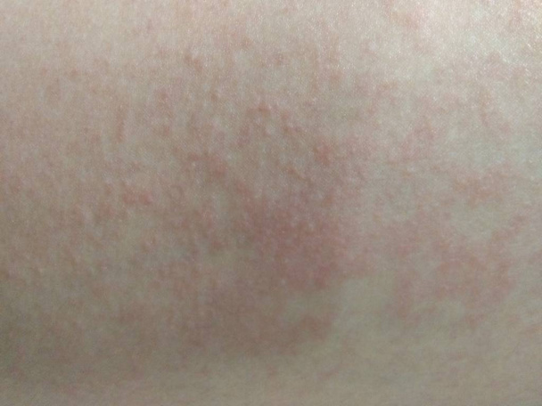
Red rash - skin reactions on the skin of women 's leg from cosmetics.
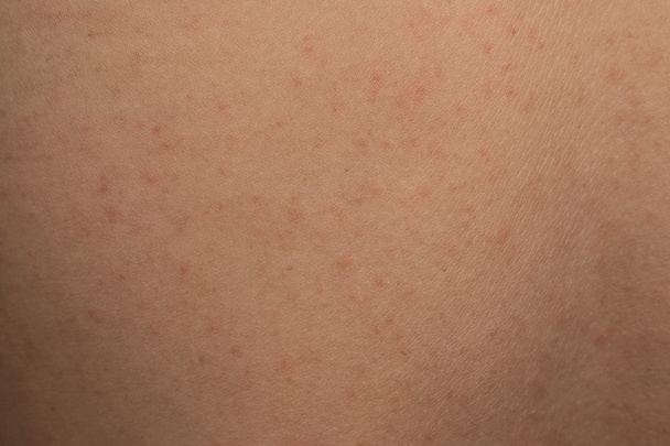
Allergic rash dermatitis skin texture of patient

Human fat body tissue under microscope view

Simple columnar epithelium is a columnar epithelium that is uni-layered. In humans, a simple columnar epithelium lines most organs of the digestive tract including the stomach, small intestine, and large intestine.

Cross section leaf of Plant under the microscope view for education.

ATP synthase 3d illustration
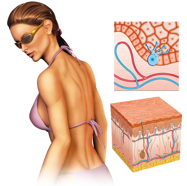
Tanned woman solarer ray

Cells of an amphibian skin with ulcer under a microscope.

Shingles is a viral infection that causes a painful rash. shingles can occur anywhere on your body, it most often appears as a single stripe of blisters that wraps around either the left or the right side of your torso.

Cross section leaf of Plant under the microscope view for education.

Cross section through cells of a seedling from a maize plant (Zea mays) under the microscope.

Section through a rosemary leaf under the microscope.

Dry Leaf Texture Abstract Background

White skin where the melanocyte secretes little melanin.

Histology of human kidney under microscope view
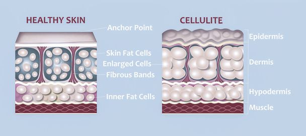
Forming of underskin cellulite illustration. Structure of normal healthy and cellulite skin. Comparison

Red acne, a rash in the chest area of a guy. closeup Unhealthy skin, dermatosis, purulent dermatitis, carbuncle, furuncle diseases of the skin and hair follicles purulent foliculitis Allergic rash dermatitis eczema skin of patient

Wart, benign skin tumor caused by human papillomavirus virus.

Photo of healthy humans skin, macro shooting

Plant cell surface of leaf under light microscope

Meso thread Lift. Young female with clean fresh skin. Beautiful woman. face and neck. Lifting by threads concept

Psoriasis is that back on white background.

Sunburns of body, closeup. Skin damaged by sun, peeling. Body care theme

3d rendering illustration of the anatomy of the skin layers. The division of each part of the structure into a layer _ dermis, epidermis, subcutaneous tissue.

Negative Allergy Skin Test on the hand of Patient.

Light photomicrograph of Onion epidermus cells whole mount seen through microscope

Blue colored leaf hairs of a mullein plant (Verbascum) under the microscope.
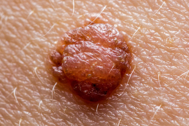
Papilloma mole on human skin, macro shot, concept of health, skin care and cancer risk

Clipped human nails on the orange background
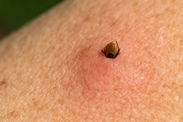
Sucking tick on human skin. Ixodes ricinus. Castor bean tick (Ixodes ricinus) bitten into pink irritated epidermis. Encephalitis, Lyme disease infection

Common wart: benign skin epithelial tumor caused by papillomaviruses.

Sunburn damaged male shoulder. The top layer of the skin peeled off. Red inflamed skin is visible.

Skin rash. Allergic reaction. Red spots on the body.

Detail of the hair of the arm of a blond boy.

Acne vulgaris in a teenager boy and closeup view of bacteria Cutibacterium acnes, formely formerly Propionibacterium acnes, associated with acne development, 3D illustration
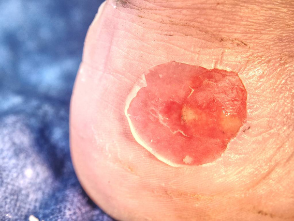
Drying and cleaning cracked bloody blister on heel. A very painful place . The heel of foot with bad skin is covered with cracks

Cross section through cells of a seedling from a maize plant (Zea mays) under the microscope.