


Filters

Stock royalty-free photos and images of Pulmonary
Discover unlimited high resolution images of Pulmonary and stock visuals for commercial use.


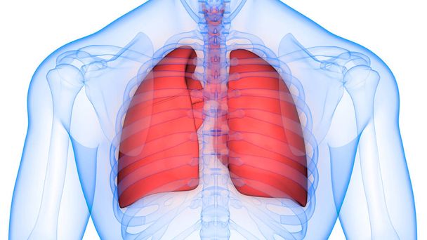
Human Body Organs (Lungs) 3D


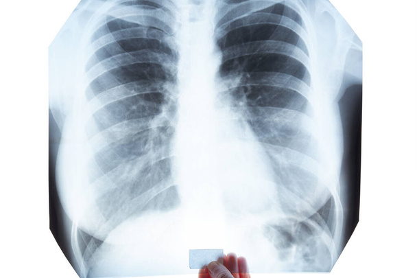
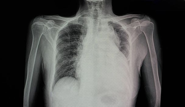
X-Ray Image Of Human Chest


Chest xray of the patient with lungs tuberculosis showing reticulonodular opacity involve both lungs from granulomatous TB infection.


X-Ray Image Of Human Chest


X-ray of the chest


X-ray of the lungs with pneumonia. Coronavirus, Covid-19.


X-ray of the lungs with inflammation with the inscription red text Coronavirus. Pandemic. Coronavirus - the viral concept of 2019, WUHAN.

X-ray of lungs in hand on white background. Isolated


A chest xray of a patient with viral pneumonia in black and white image with grey circle to identify effected area.


Abstract blurred view of blurred x-ray of human lungs.


Chest x-ray showing A 3 cm pleural based nodule at Lt upper lobe with splicated margin. Thickening of nearby pleural. Nodular infiltration at RUL. Normal heart size. differential diagnosis CA lung, TB granuloma.


Lungs ribs x-ray scan test result.


X-Ray of the chest in a patient with Active pulmonary Tuberculosis (TB) showing pneumonia and cavitation scattered in both lungs.


Male chest x-ray on black background

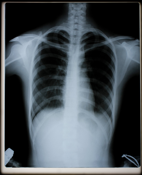
X-Ray Image Of Human Chest for a medical diagnosis

Xray film of a patient with pulmonary tuberculosis in the left upper lung (red arrow)


Chest Xray film of a patient with pulmonary tuberculosis in the left upper lung


Xray film of a patient with pulmonary tuberculosis

Chest x-ray film photos And the respiratory system for diagnosis in the treatment of diseases X-ray film images taken from the x-ray room for diagnosis of faults that require surgery for medical


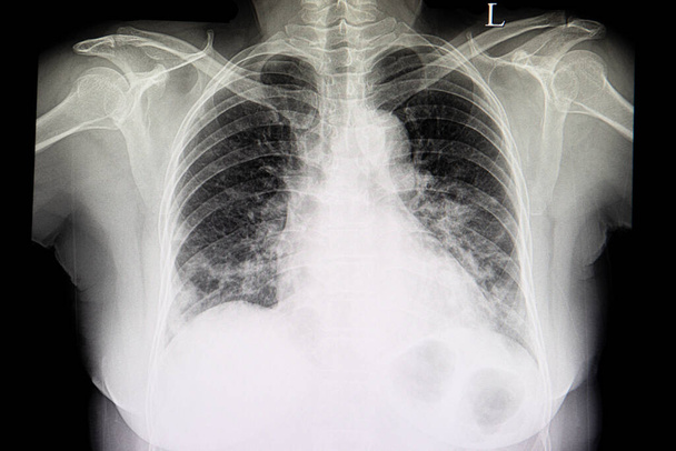
X-Ray Image Of Human male Chest


Detail of an x-ray film of lungs

A chest xray film of a patient with cardiomagaly, congestive heart failure and pulmonary edema.


Lungs ribs x-ray scan test result.

A chest x-ray of a patient with pneumonia with left lower lung infiltration

Xray film of a patient with pneumonia in his right lower lung

X-Ray Image Of Human male Chest

Anatomy collection - X-rays of lungs. Watercolor isolated organ

X-ray of a human chest or lungs radiography shot, medical technology and roentgen clinic diagnostic concept, toned

A chest x-ray film of a patient with pneumonia in his right middle lung.

Chest x-ray film photos And the respiratory system for diagnosis in the treatment of diseases X-ray film images taken from the x-ray room for diagnosis of faults that require surgery for medical

Xray film of a patient with pneumonia in his left middle lung

A chest xray film of a patient with a large lung bleb at right lower lung. The yellow marker marks the outline of the cavity. The patient could have sudden onset of pneumothorax.

Chest xray of of patient with emphysematous lungs, hilar adenopathy, and granulomatous changes of both upper lungs.

X-Ray Image Of Human Chest for a medical diagnosis. x-ray show congestive heart failure.

Chest X Ray film of a male patient with spontaneous pneumothorax with almost totally collapsed right lung. A chest drain is also seen
A doctor checks the x-ray film of the lungs and heart of a female patient in a hospital room.

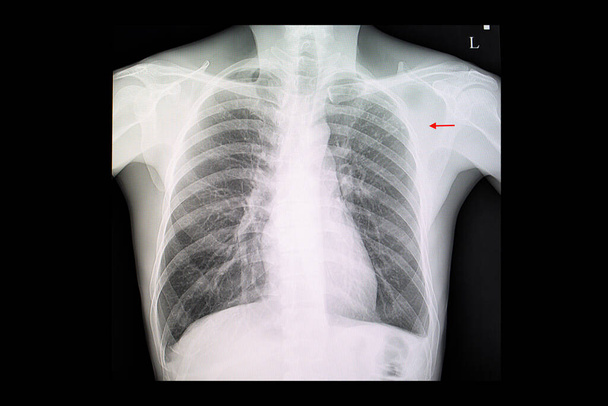
Xray film of a patient suffering car accident showing closed fracture left clavicle.

Chest radiography, lung x-ray after a covid 19 infection

A chest xray film of a patient with lung nodule in the left upper lung.

An anteroposterior X-ray of a patient diagnosed with advanced bilateral pulmonary tuberculosis. This AP X-ray of the chest reveals the presence of bilateral pulmonary infiltrate, and caving formation present in the right apical region. The diagnosi

A doctor checks the x-ray film of the lungs and heart of a female patient in a hospital room.

Chest xray film of a patient with bronchiectasis showing partial collapsed right with pleural effusion.

A chest xray film of a patient with double lumen catheter with its tips located in the right attium. The patient also has cardiomegaly.

X-Ray Image Of Human Chest for a medical diagnosis
A chest x-ray film of a patient with pulmonary tuberculosis with fibronodular infiltration at left upper lung.

A doctor checks the x-ray film of the lungs and heart of a female patient in a hospital room.

Human Respiratory System Lungs with Diaphragm Anatomy. 3D

A chest film of a patient with congestive heart failure, an enlarged heart (cardiomegaly) and pulmonary congestion is shown

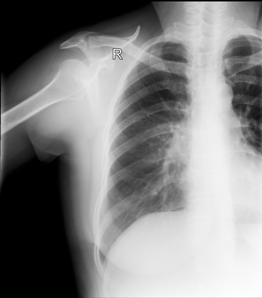
A shoulder xray film of a patient showing fractured second to to fifth rib with fracture scapular blade.

Human Respiratory System Lungs with Alveoli Anatomy. 3D

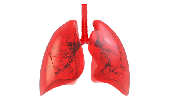
3D illustration of Lungs - Part of Human Organic.
A chest x-ray film of a female patient with cardiomegaly, pulmonary edema and right lung pleural effusion.

Human Respiratory System Lungs Anatomy. 3D illustration

Human Circulatory System heart Anatomy. 3D

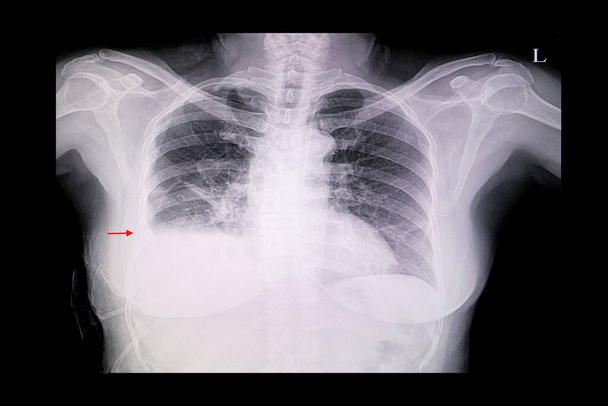
A chest xray film of a patient with dengue hemorrhagic fever in convalescence phase. A right pleural effusion is demonstrated.

Human lungs on scientific background. 3d illustration

A chest x-ray film of a female patient showing cardiomegaly and bilateral pulmonary congestion

3D illustration of Lungs - Part of Human Organic.

A doctor checks the x-ray film of the lungs and heart of a female patient in a hospital room.

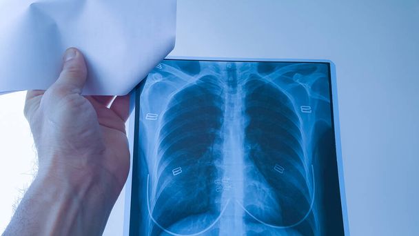
Pulmonologist with x-ray image of lungs in clinic

A chest x-ray film of a female patient with cardiomegaly and pulmonary edema.

Black and white X-ray Image of a human chest for a medical diagnosis

Human Circulatory System heart Anatomy. 3D

3D illustration of Diaphragm - Part of Human Organic.
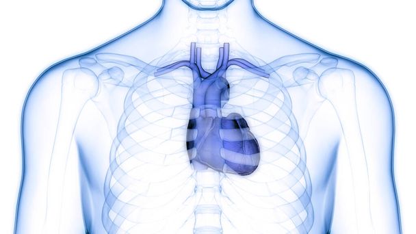
Human Circulatory System Heart Anatomy. 3D

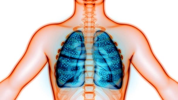
3D Concept of Human Respiratory System Lungs Anatomy
Human Respiratory System Lungs Anatomy. 3D
A chest x-ray film of a patient with cardiomegaly with left side heart failure, pulmonary edema, and pericadial effusion

Concept conceptual 3D human or man anatomy lung organ and respiratory system illustration on blue background
Lungs a Part of Human Respiratory System Anatomy X-ray 3D rendering

Human Circulatory System heart Anatomy. 3D

Human Respiratory System Lungs Anatomy. 3D

Film x-ray of chest , lung cancer from patient who love smoking cigarette , health concept

A doctor checks the x-ray film of the lungs and heart of a female patient in a hospital room.
Human Respiratory System Lungs with Alveoli Anatomy. 3D

Visual illustration of coronavirus (covid-19) infection in the lungs with 3D rendered viral particles and a chest xray film of a patient with bilateral lower lungs pneumonia.

Concept or conceptual anatomical human man 3D wireframe mesh respiratory system with lungs on blue background