


Filters

Stock royalty-free photos and images of Patologia
Discover unlimited high resolution images of Patologia and stock visuals for commercial use.


Immature leukemic cell in leukemia blood smear.

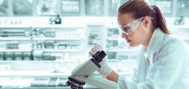
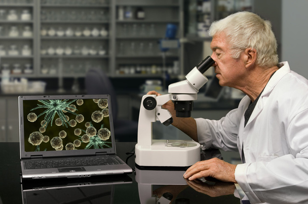
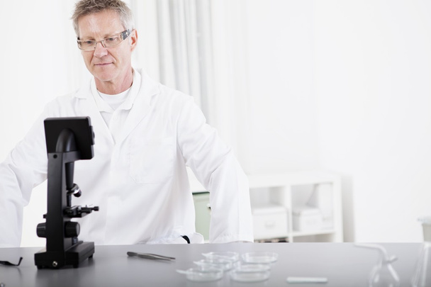
Medical Scientist Researcher Using Microscope in Laboratory, Medicine Specialist Studying and Experiment Anti Virus Pharmaceutical With Microscopic. Lab Research and Technology Health Care Concept.

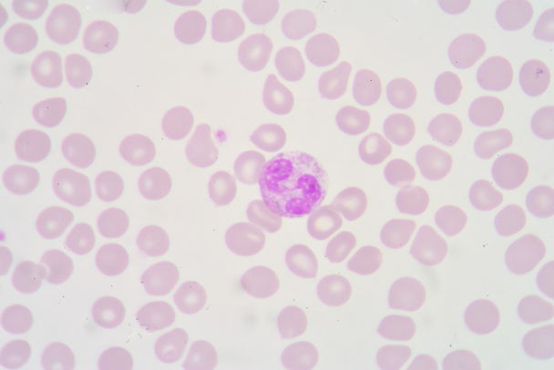
A blood smear is often used as a follow-up test to abnormal results on a complete blood count (CBC) to evaluate the different types of blood cells.


Male scientist microscoping on hi-tec fluorescent microscope.

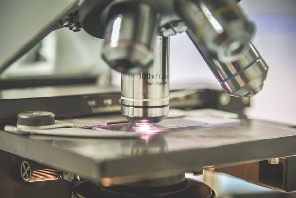
Laboratory Microscope. Scientific research background medical equipment.

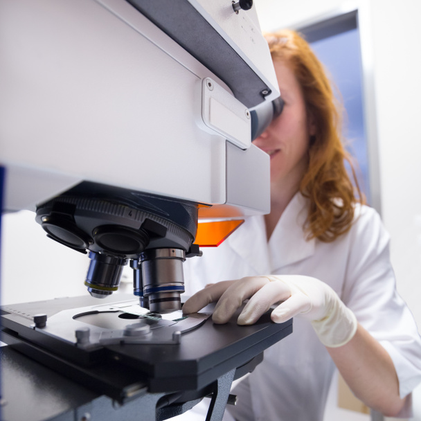
Female scientist microscoping on hi-tec fluorescent microscope. . Health care professional in hes working environment.

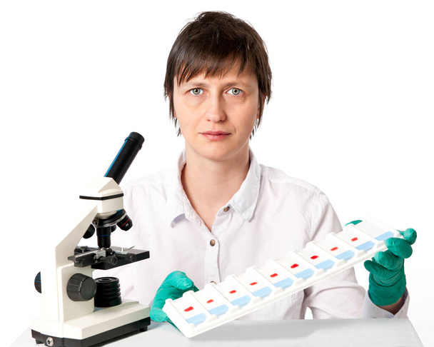
Histologist with a microscope and a tray of microscopic slides on a white background


Female scientist microscoping on hi-tec fluorescent microscope. Health care professional in hes working environment.

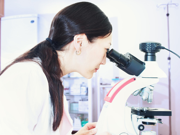
Portrait of young asian girl doctor looking to microscope in laboratory


Woman in a laboratory microscope with microscope slide in hand.toned image


Female scientist microscoping on hi-tec fluorescent microscope. . Health care professional in hes working environment.

Microscope on the table in laboratory room with laboratory equipment


Microscope on the table in laboratory room with laboratory equipment
Biologist looking through a microscope in a research lab

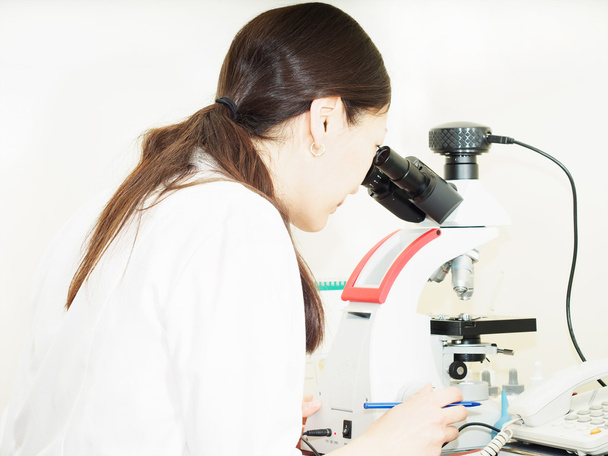
Portrait of young asian girl doctor looking to microscope in laboratory


Female scientist microscoping on hi-tec fluorescent microscope. Health care professional in hes working environment. Blue toned grayscale image.


Close-up of colorful chemical test tubes on tray in medical laboratory


Close-up on ocular, objective and slide glass of light or fluorescent microscope in research laboratory, histology or histopathology training class for medics, students or scholars in biology class.


Microscope on the table in laboratory room with laboratory equipment


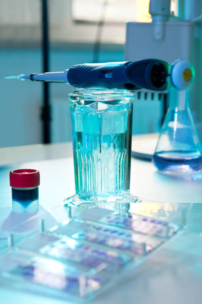
Science concept. Laboratory equipment composition. Test tubes and microscope on blue background.


Scientist in coverall clothing is examining coronavirus sample in a laboratory.The researcher is using a loupe.

Test finds the first signs of a tumor in the blood, Based on DNA analysis for the 'super-early' diagnosis

Laboratory with microscope in the foreground


Men in a laboratory microscope with microscope slide in hand.


Female professional medical tech works with microscope in research laboratory, histology or histopathology. Microscopic analysis of cell cultures to develop vaccine for coronavirus to fight covid-19.


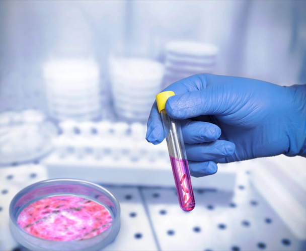
Hand in glove, medical laboratory scientist hold blood sample in test tube. Stand with more blood, urine samples for medical analysis in test lab. Laboratory study of samples for novel coronavirus.


Portrait of young asian girl doctor looking to microscope in laboratory


Scientist examines sample of coronavirus in laboratory, conceptual image


Portrait of young asian girl doctor looking to microscope in laboratory


Scientist adjusting knobs of a light microscope

Test tube with sample in pre enrichment medium Buffered Peptone Water collected from animals

Rotary Microtome Section for diagnosis in pathology make microscope slide pathology. Human tissue equipment.

Laboratory doctor sets test tubes with blood sample into centrifuge
Portrait of male medical or scientific researcher with microscope in a laboratory
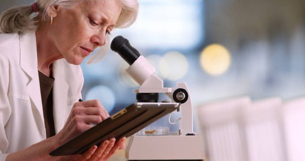
Mature female scientist takes notes on tablet computer and uses microscope
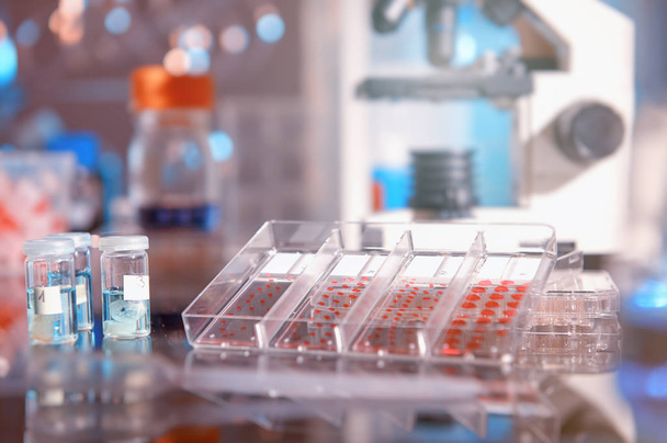
Scientific background with modern histopathology tools: stained microscopic slides of patient tissue, fixed tissue samples and a microscope. Space for your text. This image is toned.

Blood analysis, clinical or medical testing and phlebotomy concept theme with close up on doctor hand wearing blue latex gloves and holding a test tube isolated on white background

Staphylococci bacteria, blood test tube samples

Scientific sampling of eggs in poor condition, analysis of avian influenza in humans, conceptual image

Hand in glove, medical laboratory scientist hold blood sample in test tube. Stand with more blood, urine samples for medical analysis in test lab. Laboratory study of samples for novel coronavirus.

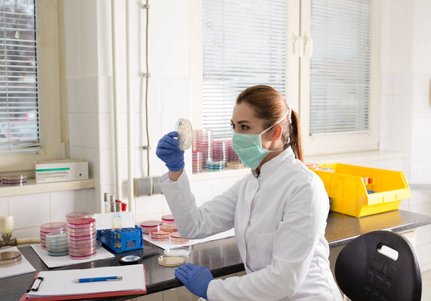
Biologist with safety workwear holding petri dish with bacteria in laboratory
Histochemical staining of tissue samples

Laboratory Glassware containing different colored liquids against a neutral background
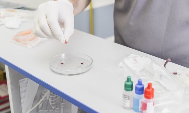
Hands of the doctor doing the analysis and a blood sample at a table with gloves


Microscope with slide sample and petri dish medical microbiology science laboratory blue banner background

Laboratory equipment composition. Science concept.

Histology test lab in hospital. Hands in gloves, tech loads slide glasses with patient biopsy sections. Staining of lung tissue. Anatomic analysis of biopsy samples from patients with viral pneumonia.

Hand of a nurse with a yellow tube with a urine sample

Vacutainer Plastic Serum Tube used for evacuated sterile blood collection tube for serum analysis
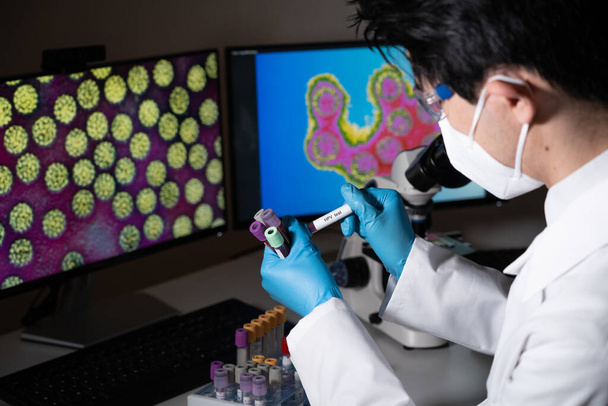
Papillomavirus test male doctor


Laboratory equipment composition. Science concept.
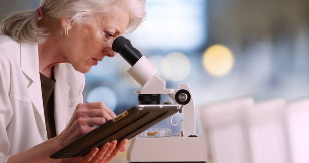
Mature female scientist takes notes on tablet computer and uses microscope

Hands of the doctor doing the analysis and a blood sample at a table with gloves


Doctor Collecting a nasopharyngeal nose and throat swab

Laboratory interior. Laboratory equipment glass beakers and microscope. Science concept.

Laboratory interior. Laboratory equipment glass beakers and microscope. Science concept.

Microscope objectives toned in blue, copy space on the right


Anatomic pathology testing. Histology test lab in hospital. Hands in gloves arrange patient biopsy sections in plastic tubes. high throughput automatic histological staining

Rotary Microtome Section for diagnosis in pathology
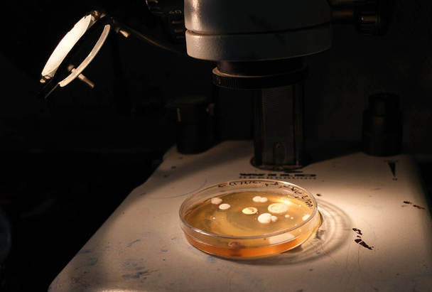
Basra, Iraq - april 30, 2021: photo of fungi growth on agar media in petri dish

Basra, Iraq - april 30, 2021: photo of fungi growth on agar media in petri dish

Basra, Iraq - april 30, 2021: photo of fungi growth on agar media in petri dish