


Filters

Stock royalty-free photos and images of Maligne
Discover unlimited high resolution images of Maligne and stock visuals for commercial use.

Young sad girl, cancer patient looking through hospital window.

Young cancer patient standing in front of hospital window.

Virus and bacterium medical microscopic bacteria cell

Businessman looks for the virus

Oral Cancer Showing Cancerous Growth And Sickness

Concept of businessman that searches virus

Prostate Adenoma - Printed Diagnosis with Blurred Text. On Background of Medicaments Composition - Red Pills, Injections and Syringe

Oncology - Inscription on Red Road Sign on Sky Background.

Leukosis - Printed Diagnosis with Blurred Text. On Background of Medicaments Composition - Red Pills, Injections and Syringe

Ovarian Cancer - Printed Diagnosis with Red Pills, Injections and Syringe. Medical Concept with Selective Focus

Cancer Treatment - Printed Diagnosis with Blurred Text on Green Background and Medical Composition - Stethoscope, Pills and Syringe. Medical Concept. 3D Render.

Handwriting text writing Hpv. Concept meaning Human Papillomavirus Infection Sexually Transmitted Disease Illness written Sticky note paper within Paper Balls Plain background Wooden Toy

HPV - Printed Diagnosis on Orange Background and Medical Composition - Stethoscope, Pills and Syringe. Medical Concept. Blurred Image. 3D Render

Handwritten Diagnosis Thyroid Cancer in the Disease Extract. Medicaments Composition of Heap of Pills, Blister of Pills and Bottle of Tablets. 3D Render.

Handwritten Diagnosis Sarcoma in the Extract From the History of Disease. Medicaments Composition of Heap of Pills, Blister of Pills and Bottle of Tablets. 3D Render.
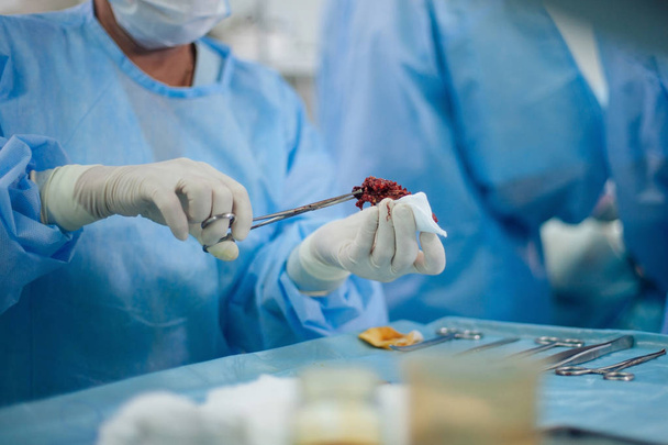
Malignant tumor removed holden by nurse in surgical clamp close-up
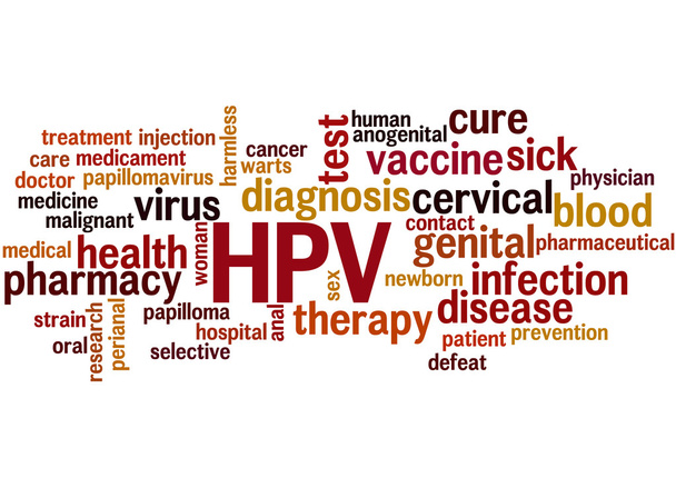
HPV - Humani Papilloma Virus, word cloud concept on white background.

Genital warts, word cloud concept on black background.

Cancer Treatment word cloud concept on white background.
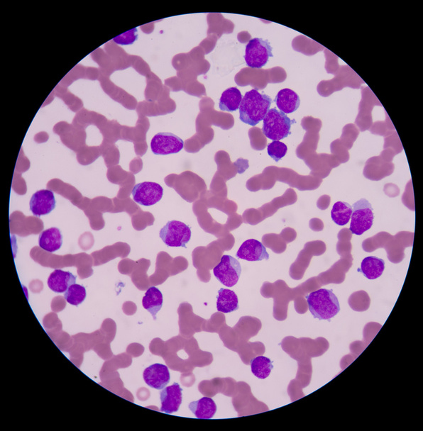
Mix shape white blood cells, concept funny wbc. blood smear is often used as a follow-up test to abnormal results on a complete blood count (CBC) to evaluate the different types of blood cells.
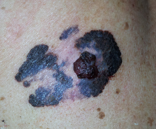
Malignat melanoma, cancer of skin

A mum applying a protect sunscreen on the face of her cute young girl
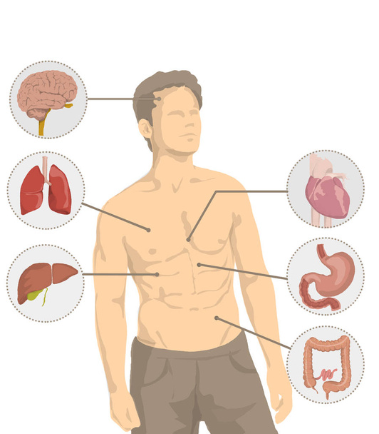
Illustration of shirtless man with the main organs of the human body, heart, brain, liver, intestine, stomach, lungs
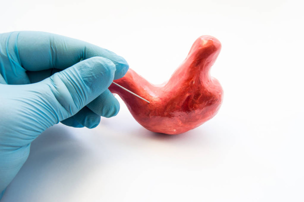
Concept of stomach puncture or gastrointestinal perforation. Hand of surgeon pierces wall of model of human stomach for therapeutic purposes or for biopsy tissue analysis by histology or cytology

Sick woman desperate in pain in a hospital.

Stomach adenocarcinoma, gastric cancer, 3D illustration and light micrograph
Thyroid cancer cells, 3D illustration
Bladder transitional cell carcinoma, light micrograph, photo under microscope
Melanoma or skin cancer. layers of the human skin.

Woman looking suspicious mall on her skin
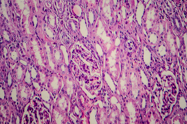
Wilms tumor, or nephroblastoma, light micrograph, photo under microscope
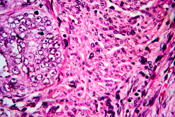
Endometrial adenocarcinoma, light micrograph, photo under microscope

Cancer Diagnosis. Medical Concept with Composition of Medicaments - Red Pills, Injections and Syringe. Selective Focus. 3D Render
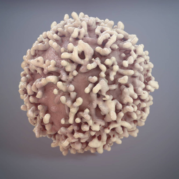
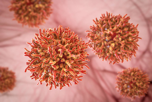
Cancer Cell on grey background - 3D Rendering
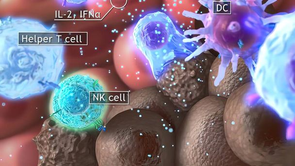
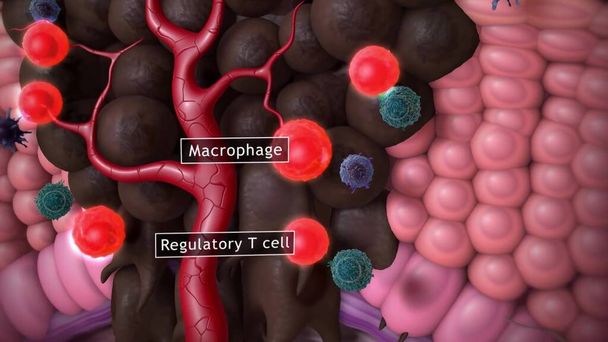
3D medical illustration cytotoxic T cell and Helper T cell destroys the stress-related molecule

Stomach adenocarcinoma, gastric cancer, 3D illustration and light micrograph
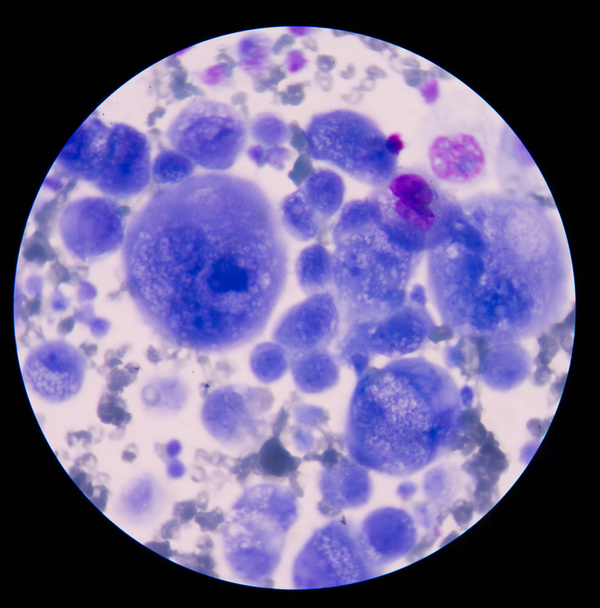
Malignant cells with multiple nuclei and prominent nucleoli.

Young cancer patient standing in front of hospital window.
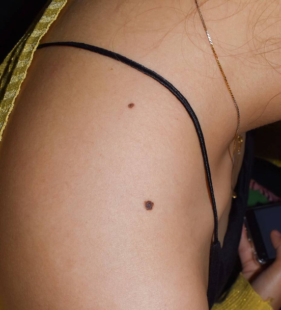
Junctional nevus or double moles at shoulder of Southeast Asian, Myanmar young woman. It is found at border between epidermis and dermis layers of skin. These moles may be colored and slightly raised.

Young woman with moles on white background

ORTHOPEDICS CONSULTATION MAN

A woman's hand hold a gray ribbon of awareness of Parkinsons disease. As a symbol of Parkinsons Day.

Doctor examining woman skin for melanoma

CG medical illustration - malignant disease or virus infection

Dermatologist examining moles of patient on light background
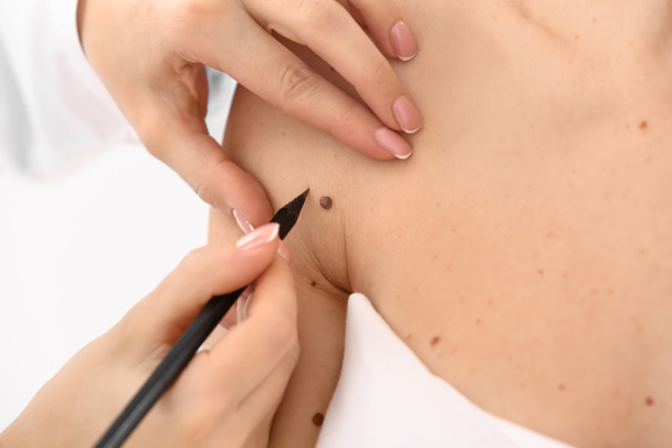
Dermatologist applying marks onto patient's skin before moles removal, closeup
3d computer illustration of a dendritic cell. They areantigen-presenting cells of the immune system. Their main function is to process antigen material and present it on the cell surface to the T cells of the immune system. They are messengers betwe

Dendritic cells present antigens (green) to lymphocytes through their membran bound MHC-molecules (violet). CD4 molecules (light blue) bind to other portions of the MHC, strengthening the interaction.

Cancer cells express PD-L1 (orange) proteins on their surface to trick the immune system. The interaction of PD-L1 with PD-1 of T-cells leads to a down-regulation of T-cells. The antibody (yellow) blocks this interaction.

Young woman with moles on white background

Metaphor about the worst disease because of smoking.
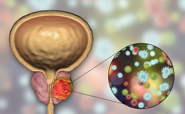
Conceptual image for viral ethiology of prostate cancer. 3D illustration showing viruses infecting prostate gland which develops cancerous tumor
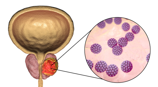
Conceptual image for viral ethiology of prostate cancer. 3D illustration showing Human Papilloma Viruses HPV infecting prostate gland which develops cancerous tumor

Angioma on the skin. Red moles on the body. Many birthmarks

Turquoise or Robin egg blue ribbon on human hand old aged background raising awareness on Bone tumor, Addiction recovery
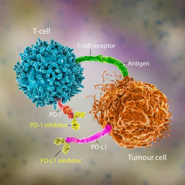
Immune checkpoint inhibitors in cancer treatment, 3D illustration. Inhibitors of PD-1 receptor and PD-L1 prevent the tumour cell from binding to PD-1 and enable the T cell to remain active

Old man visiting male doctor dermatologist

Dermatologist examines the patient's mole on the back

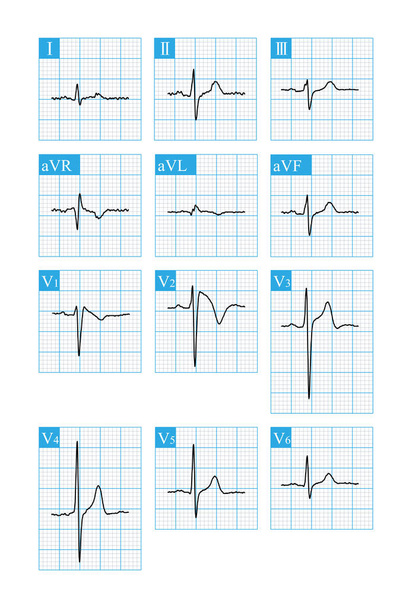
X-rays

Cancer Treatment , Doctor writing on transparent screen
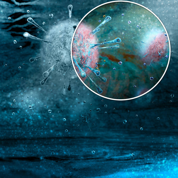
Virus, genetic code, bacteria and microorganisms seen under a microscope. New viruses

Young depressed cancer patient standing in front of hospital window.

Skin Cancer - Medical Concept on Blue Background with Blurred Text and Composition of Eyeglasses. 3D Rendering.

Old man visiting male doctor dermatologist
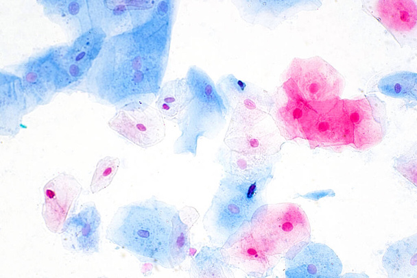
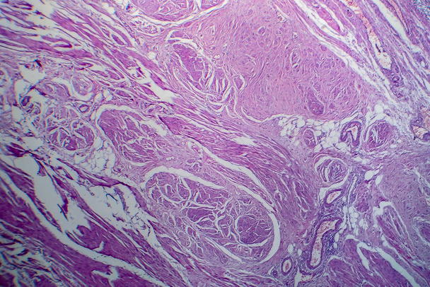
Squamous epithelial cells under microscope view for education histology. Human tissue.

Young depressed cancer patient standing in front of hospital window.

Primary myelofibrosis (PMF) cells in blood flow - closeup view 3d illustration
Meningioma (brain cancer) tumor in the brain tissue - 3d illustration closeup view

Dermatologist examining mole on young man's back against blue background, closeup

Dermatologist examining mole on young man's shoulder against pink background, closeup

The dermatologist examines the moles or acne of the patient with a dermatoscope. Prevention of melanoma

3D render of two connected puzzle pieces with the words melanoma and genetics, symbolizing the link between genes and the deadly skin cancer.

A man in beige jeans at the level of the genitals , holding a ripe and rotten apple. Disease for men. The concept of protection of sexually transmitted infections. Copy space. Blue tint.

Dermatologist examining patient in clinic, closeup

Uterine cancer, 3D illustration showing malignant tumor in the female uterus and close-up view of a cancer cell

Man suffering from stomach pain and injury isolated white background. Health care and medical concept.

A man at the level of the genitals , holding a ripe and rotten apple. Male suffering with pain in the urogenital system. Disease for men. The concept of protection of sexually transmitted infections. Close up.

Sick senior woman suffering from appendicitis,severe abdominal pain,female patient with a stomach ache sit in a hospital bed,problems with liver disease or irritable bowel syndrome,colorectal cancer

One person is answering question about breast screening.

Human skin with moles, closeup. Concept of skin cancer

Young man visiting old doctor dermatologist

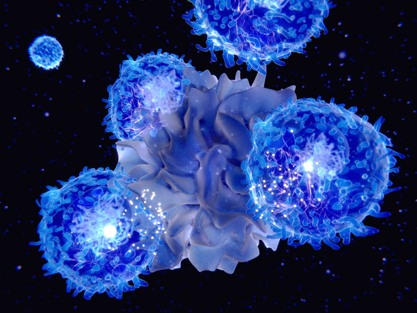
T-lymphocytes attacking cancer cell, 3D illustration isolated on white background with clipping path. Anticancer immunity and treatment concept

T-lymphocytes attacking cancer cell, 3D illustration with clipping path. Anticancer immunity and treatment concept

Cancer cells, malignant cells, scientific 3D illustration

Ovarian cancer, 3D illustration showing malignant tumor in the left ovary

Ovarian cancer, 3D illustration showing malignant tumor in the left ovary

Electrocoagulation and laser cosmelotogy. Removal of birthmark from female face. Dermatologist surgeon using a professional electrocautery for removing mole. Comparison of results before and after operation.
A 36 year old man survived CPR after sudden syncope. The electrocardiogram was suggestive of Brugada syndrome type 1. Implantation of ICD therapy.

Dermatologist examining moles on young woman's belly in clinic, closeup
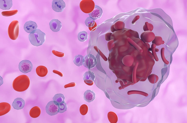
Auer rods (or Auer bodies) in acute Hypergranular Promyelocytic Leukemia (APL) - 3d illustration closeup view

During stage III endometrial cancer, the tumor spreads outside of the uterus. 3D rendering

Stage II endometrial cancer is characterized by tumor spread to the uterine cervix. 3D rendering
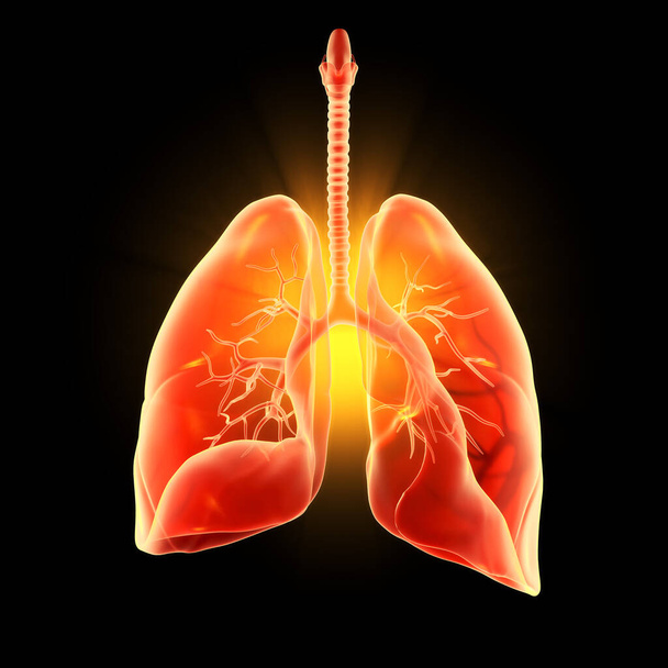
Illustration showing highlighted human lungs, pneumonia, 3D illustration

A medical consultation at the removal of nevus

Handwriting text Cancer Treatment. Conceptual photo The analysisagement of medical care given to a cancer patient

Asian lady woman patient have abnormal enlargement of thyroid gland Hyperthyroidism (overactive thyroid) at the throat : healthy strong medical concept
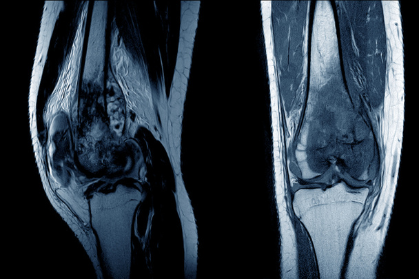
MRI Knee joint (AP,LATERAL)Views a female 15 year old showing Osteosarcoma.Medical healthcare concept.
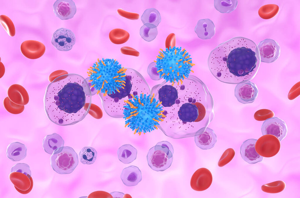
CAR T cell therapy in Multiple myeloma (MM) - isometric view 3d illustration

A 3D scientific illustration showcasing a human body with transparent skin, revealing a tumor in his thyroid gland, along with a micrograph image of papillary thyroid carcinoma.

Dermatologist examining patient on color background